For thousands of years, humanity looked upon the Moon in awe and terror, in light of its perceived evil influence. Then Johannes Hevelius, the son of a wealthy Polish brewer, built a homemade telescope in what is now Gdansk and sat down nightly to scrutinize its extraterrestrial world. In 1647, he published the first book of maps of the Moon.
Now, Tomasz Nowakowski, a 40-year-old neuroscientist who…
For thousands of years, humanity looked upon the Moon in awe and terror, in light of its perceived evil influence. Then Johannes Hevelius, the son of a wealthy Polish brewer, built a homemade telescope in what is now Gdansk and sat down nightly to scrutinize its extraterrestrial world. In 1647, he published the first book of maps of the Moon.
Now, Tomasz Nowakowski, a 40-year-old neuroscientist who grew up in the same city as Hevelius, is leading the creation of thefirst draft map of the human brain during its developmental stages — from embryo to adulthood. The researcher says he feels like one of those early cartographers. “Humans would never have landed on the Moon if we hadn’t had a map of the lunar surface. All the great advances and achievements in history began with the creation of accurate maps,” he tells EL PAÍS.
Nowakowski is one of the leaders of the BRAIN Initiative, a U.S. project launched by president Barack Obama in 2013 to map the human brain — an effort that has already amassed an enormous budget of €4.5 billion ($5.21 billion). “The brain, the origin of our thoughts, ideas and imagination, continues to be the most important unexplored object. To understand it, we must start by understanding its list of components,” says Nowakowski, from the University of California, San Francisco.
That’s a monumental job. During pregnancy, a single cell — the egg fertilized by the sperm — multiplies, and starting at the third week, the development of a rudimentary nervous system begins, a process that culminates in 86 billion neurons and trillions of connections between them. In this unimaginable choreography inside a fetus’s skull, some cells take alternative paths. Nowakowski cites estimates that 15% of children and adolescents live with a neuro-developmental disorder like autism, schizophrenia or attention deficit hyperactivity disorder.
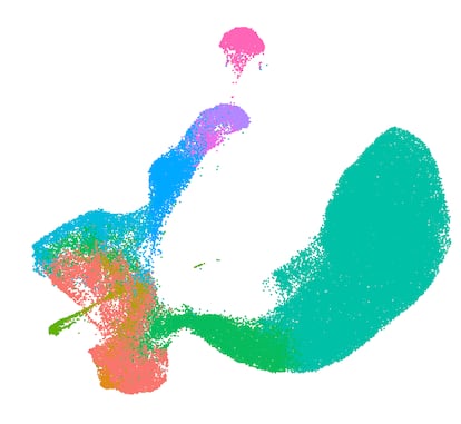
The BRAIN Initiative’s international consortium is now using the latest technologies, capable of analyzing which genes are active in each cell, to draw up the first draft of a dynamic map of the developing brain. “Human tissue can be obtained from surgical procedures or from post-mortem brains that would normally be discarded. If the cells are isolated from the tissue quickly enough, they can be cultured in vitro for a few hours or, in some cases, for several days. This gives us a unique opportunity to study developmental processes in humans,” Nowakowski says. So-called pluripotent stem cells, obtained from surplus embryos from fertility clinics or reprogrammed adult cells, now also make it possible to mimic the early stages of brain formation in a lab.
These recent results are first steps towards understanding the specific moments during pregnancy in which there is the highest risk of brain tumors or neurological development anomalies. Genes that are implicated in mental disorders like autism and schizophrenia, explains Nowakowski, are activated at greater intensity at the end of gestation, precisely at the stages that differ most from the functioning of mice and other laboratory animals. It is essential to have our own atlas for understanding how the human cerebral labyrinth is formed.
The brain contains thousands of cell subtypes, each highly specialized in its functioning: neurons for thinking, astrocytes that play supporting roles, oligodendrocytes that function as the insulating layer of neural cables, and microglia that clean waste from the nervous system. The consortium has found that during fetal development, cells are extraordinarily flexible in terms of their identity, allowing them to become other cell types in the adult brain. That flexibility is also their weakness. Researchers have detected a type of progenitor cell, present in the second trimester of pregnancy, that can generate neurons as well as oligodendrocytes and astrocytes. An incurable brain cancer called glioblastoma has cells similar to this progenitor, which offers a clue as to the origin of the tumor.
The BRAIN Initiative Cell Atlas Network announced the results of a half-dozen studies, published in Nature* *this week. The first line of the article recalls the work of Spain’s Santiago Ramón y Cajal, the man who, armed with a microscope and chicken cerebellums, presented the first objective evidence in 1888 that the nervous system is organized into individual cells. “Almost all modern neuroscience is based on the concepts proposed by Cajal,” says Nowakowski. “He was a visionary, even in areas that we still don’t know how to study. I am convinced that his vision will continue to resonate for many years to come,” adds the University of California professor.
Spanish neuroscientist Rafael Yuste was there in the early days of the BRAIN Initiative. He says that one day in September 2011, at the British mansion Chicheley Hall, two dozen experts on the brain and the study of structures measuring millionths of a millimeter across assembled to discuss possible ways of collaborating. Yuste stood up and delivered comments that led to much debate: he proposed analyzing all neurons, one by one. Examining only a few, he said, was like trying to watch TV by looking at a single pixel. Amid voices claiming that the idea was unfeasible, U.S. geneticist George Church, who had been in favor of a comprehensive project examining human DNA since 1984, stood up and declared that in science, “nothing is impossible.” The White House adopted the proposal and, in early 2013, Obama solemnly announced the official start of the project. “As humans, we can identify galaxies light years away, we can study particles smaller than an atom. But we still haven’t unlocked the mystery of the three pounds of matter that sits between our ears,” the president declared.
Yuste, a Columbia University professor, is excited by the initiative’s newest results. “This atlas of developmental cellular types is essential, not just for the scientific understanding of how the brain develops — which is absolutely fascinating when one keeps in mind that it assembles and organizes itself with no external direction — but also, it is fundamental information towards understanding the alterations and pathologies that take place during pregnancy in and in the first stages of life,” he says. “These results show how sustained investment in the development and application of new methodologies is of crucial importance in science and medicine,” adds Yuste, who is working on plans for Spain NeuroTech’s NeuroTech National Center.

Two years ago, neuroscientist Hongkui Zeng’s team presented the most complete mapping of an adult rodent’s brain: an organ the size of a pea with barely 70 million neurons and 5,300 kinds of cells. Zeng and his colleagues from the Seattle-based Allen Institute for Brain Science have now turned their focus to cells that are essential to the functioning of the nervous system: GABAergic inhibitory neurons, which act as the brain’s break, diminishing its activity to facilitate the transmission of information. These neurons, according to the researchers, continue developing after birth, particularly in the areas of the brain related to learning, emotions and decision-making. “This means there could be a longer period than previously thought for intervening and helping the brain to reorganize, especially in the case of children with development disorders,” says the Allen Institute in a statement.
Neuroscientist Giullermina López Bendito speaks of a “qualitative leap” in her field of study. “Until now, we had cell atlases primarily of adult brains, which provided a static view of cell identity. This collection of articles turns that snapshot into a moving film: it reconstructs the temporal progression and cell lineages that give rise to the developing brain,” says the researcher from the Institute of Neurosciences in Alicante, Spain.
López Bendito leads a laboratory that is looking to understand the dense network of connections between brain cells. She points out that the consortium, in which she did not participate, has mapped different species of mammals, from mice to humans, identifying specific characteristics of humans and other primates, such as the longer period of new neuron generation and the diversification of the cerebral cortex after birth. “These temporal differences could be at the root of both human cognitive abilities and our greater vulnerability to genetic mutations or environmental alterations during development,” notes the neuroscientist.
Alone with his microscope in 1888, Santiago Ramón y Cajal undertook the impossible mission of mapping the brain, millimeter by millimeter. His work culminated in 1904 with the publication of *Texture of the Nervous System of Man and the Vertebrates, *a colossal 1,800-page work that came with a warning that the completion of the edifice of neurology will still require the work of many centuries.
Tomasz Nowakowski is more optimistic. “I don’t think we are centuries away, or even decades. I have been impressed by the speed with which artificial intelligence has advanced, especially in recent years, to emulate certain cognitive processes through computational models,” reflects the researcher. “I believe we are rapidly approaching a point where theory and modeling will make predictions about which cells and molecules are essential for the structure and function of the brain. And we will have the technologies necessary to test those predictions.”
*Sign up for our weekly newsletter to get more English-language news coverage from EL PAÍS USA Edition *