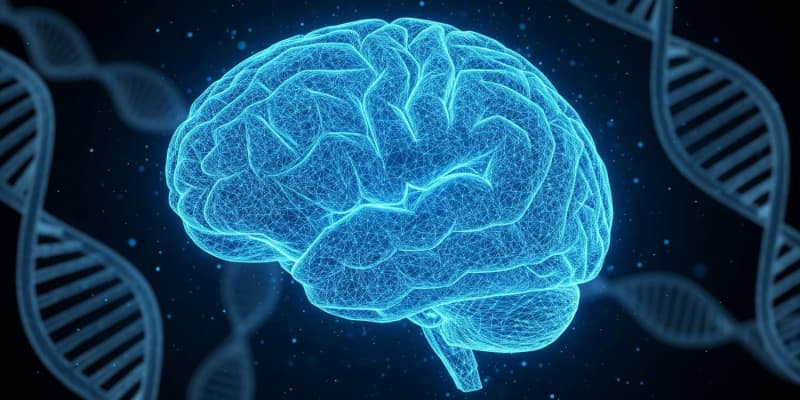
Researchers have created a detailed map of chemical modifications to DNA that occur in the human brain from the earliest stages of fetal development through old age. A new study published in Cell Genomics reveals that the most significant of these changes happen before birth and are located near genes linked to conditions like autism and schizophrenia.
The human brain cortex, the wrinkled outer layer responsible for thought, memory, and behavior, undergoes a period of intense and complex construction before birth. This process relies on genes being switched on and off in a precise, time-sensitive sequence.
Epigenetic change…
Researchers have created a detailed map of chemical modifications to DNA that occur in the human brain from the earliest stages of fetal development through old age. A new study published in Cell Genomics reveals that the most significant of these changes happen before birth and are located near genes linked to conditions like autism and schizophrenia.
The human brain cortex, the wrinkled outer layer responsible for thought, memory, and behavior, undergoes a period of intense and complex construction before birth. This process relies on genes being switched on and off in a precise, time-sensitive sequence.
Epigenetic changes, which are chemical tags that attach to DNA and direct gene activity without altering the genetic code itself, are fundamental to this orchestration. A team of researchers at the University of Exeter sought to understand the dynamics of one key epigenetic mechanism, DNA methylation, across the entire human lifespan to build a clearer picture of how the cortex develops.
To create this comprehensive timeline, the scientists analyzed DNA methylation in the cortex tissue of nearly 1,000 donated human brains. The samples spanned a remarkable age range, from just six weeks after conception to 108 years of age. Using a technology that can measure methylation levels at hundreds of thousands of specific sites across the genome, they identified which sites changed with age. They first looked at the whole tissue, then developed a method to zoom in on specific cell populations to see if changes were uniform across the brain’s different cell types.
The team’s initial analysis revealed that the prenatal period is a time of extraordinary epigenetic activity. They identified more than 50,000 specific locations on the DNA where methylation levels changed dramatically during early and mid-gestation. Many of these changes were not linear, meaning they did not happen at a steady rate. Instead, they appeared to accelerate, slow down, or level off at distinct developmental time points, suggesting they mark important biological transitions in the construction of the cortex.
When the researchers compared these early-life changes to those happening after birth, they found a striking difference. The vast majority of the methylation patterns established before birth remained relatively stable throughout the rest of life. Only a very small percentage of these sites continued to change significantly with age postnatally. This finding indicates that the prenatal window is a unique period of extensive epigenetic remodeling that largely sets the stage for future brain function.
The scientists noticed that these dynamically changing DNA sites were not spread randomly. They were depleted in some genomic regions but concentrated in others, particularly in areas known to regulate gene activity. These sites were also enriched in parts of the genome that are accessible and active during the development of specific brain cells, especially excitatory neurons, which are the brain’s primary signaling cells. This suggests that the methylation changes are tied to the activation of genetic programs needed to build a functional cortex.
Recognizing that the brain is composed of many different cell types, the team wanted to know if these epigenetic changes were happening in all cells or were specific to certain ones, like neurons. Analyzing the cortex as a whole tissue can mask cell-type-specific patterns. The standard protein marker used to identify mature neurons, called NeuN, did not work reliably on the very immature neurons found in the early fetal brain. The researchers established a new protocol using a different marker, a protein called SATB2, which is active in developing excitatory neurons.
Using a method that sorts cell nuclei based on fluorescent labels, they separated the brain tissue into two populations: one enriched with developing neurons and another containing other brain cell types. They then analyzed the DNA methylation in each group separately. This approach showed that neurons begin to establish their unique epigenetic signature very early in development. The majority of the developmental changes seen in the whole cortex tissue were, in fact, being driven by shifts occurring within this developing neuron population.
The analysis also revealed distinct developmental trajectories between the cell types. Some methylation sites changed only in the neuron-enriched group, while others were specific to the non-neuronal cells. For example, the sites changing dynamically only in the developing neurons were located in active genomic regions specific to excitatory neurons. Sites changing only in the other cell populations were, by contrast, enriched in active regions associated with astrocytes, a type of supportive brain cell.
Finally, the researchers investigated whether these dynamic epigenetic regions were relevant to neurodevelopmental conditions. They examined lists of genes that have been strongly associated with autism and schizophrenia through genetic studies. They found that these genes were significantly more likely than other genes to be located near the DNA sites that undergo methylation changes during fetal development.
This association was even stronger when they looked specifically at the changes happening within the developing neurons. The findings support the long-standing hypothesis that the origins of these conditions may lie in disruptions to early brain development.
The study does have some limitations. Access to brain tissue from later stages of pregnancy was restricted, though the data suggest that the most dramatic changes had already occurred by mid-gestation. The technology used, while extensive, only covers a fraction of all possible methylation sites in the genome.
Future research using whole-genome sequencing could provide an even more complete view. The method was also unable to distinguish between DNA methylation and a related modification, DNA hydroxymethylation, which is also common in the brain. Despite these constraints, the study provides a foundational resource for understanding the epigenetic processes that shape the human brain.
The study, “Cell-type-specific DNA methylation dynamics in the prenatal and postnatal human cortex,” was authored by Alice Franklin, Jonathan P. Davies, Nicholas E. Clifton, Georgina E.T. Blake, Rosemary Bamford, Emma M. Walker, Barry Chioza, Martyn Frith, APEX Consortium, Youth-GEMs Consortium, Joe Burrage, Nick Owens, Shyam Prabhakar, Emma Dempster, Eilis Hannon, and Jonathan Mill.